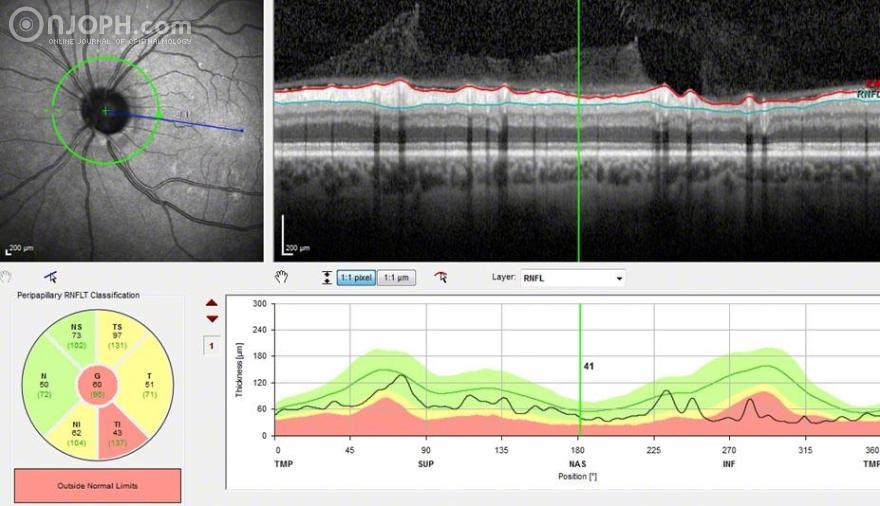
OCT Circular Scan: decreased retinal nerve fiber layer thickness RNFL at the inferotemporal sector.
Patient: 68 years of age, female, BCVA 1.0 at OD, 0.8 at OS, IOP 12/9 mmHg.
Ocular Medical History: Unilateral increased intraocular pressure in OS since 2013, filtrating surgery in OS in 2014.
General Medical History: arterial hypertension.
Purpose: to present optic pit in glaucomatous optic nerve atrophy.
Methods: Colour Photography Posterior Pole, Optical Coherence Tomography, Visual Field.
Findings:
Colour Photography Posterior Pole: thinned rim and optic disc pit at the inferotemporal periphery of the optic disc.
Optical Coherence Tomography at 5 o’clock: yellow line indicates the scanned optical coherence tomography (OCT) lines, B-scan image illustrates the no hyporeflective space indicating an optic disc pit.
Optical Coherence Tomography at 6 o’clock: yellow line indicates the scanned optical coherence tomography (OCT) lines, B-scan image illustrates the hyporeflective space (arrowhead) at the location of optic disc pit.
Optical Coherence Tomography at 7 o’clock: yellow line indicates the scanned optical coherence tomography (OCT) lines, B-scan image illustrates the hyporeflective space (arrowhead) at the location of optic disc pit.
Optical Coherence Tomography at 8 o‘clock: yellow line indicates the scanned optical coherence tomography (OCT) lines, B-scan image shows no hyporeflective space indicating an optic disc pit.
OCT Circular Scan: decreased retinal nerve fiber layer thickness RNFL at the inferotemporal sector.
Visual Field: parafoveal scotoma in the superior hemifield corresponding to the decreased RNFL thickness at the inferotemporal sector.
Discussion:
Choi et al. (1) reported that optic disc pits had a diverse structure according to the alteration of the lamina cribrosa or prelaminar tissue. Optic disc pit is often associated with a corresponding scotoma. Choi et al. (1) suggested, that a compliant lamina cribrosa or prelaminar tissue may become susceptible to the damaging effects of increased IOP values. The ongoing insult to the optic nerve may then influence the lamina cribrosa tissue or prelaminar tissue, exhibiting the appearance of alteration in the lamina cribrosa or or prelaminar tissue. Subsequently, the axons passing through the altered lamina cribrosa and or prelaminar tissue are likely to be damaged by loss of structural support, loss of nutrient supply from laminar capillaries, or loss of metabolic support from astrocytes.
Literature:
(1) Choi YJ, Lee EJ, Kim BH, Kim TW. Microstructure of the optic disc pit in open-angle glaucoma. Ophthalmology. 2014 Nov;121(11):2098-2106.